

Bristol Knee Clinic

| + home | |
| + news | |
| + research | |
| + patient information | |
| + the clinic | |
| + the surgeon | |
| + sport physiotherapy | |
| + sports advice | |
| + medico legal | |
| + products | |
| + resources | |
| + contact | |
| + maps | |
| + directions | |
| + site map |
The Bristol Knee Clinic |
Arthroscopy Of The Knee - Indications / Contra-indications
Anatomy / Ailment Detail 
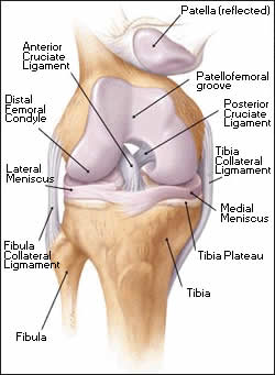
The knee comprises the joint between the femur and the tibia but also the joint between the patella and the front of the femur. Between the femur and the tibia sit two crescentic cartilages or menisci. These fibro-cartilaginous discs dissipate the compressive forces between across the knee and thereby avoid excessive loading, wear and damage. The ligaments around the knee stabilise the knee. They include the collateral ligaments; medial and lateral, lying either side of the knee and the cruciate ligaments, anterior (ACL) and posterior cruciate ligament (PCL), lying within the joint. The fibrous capsule, which surrounds the knee, completes the stability of the joint.
The two most important groups of muscles supporting the knee are the quadriceps muscle, which is the large bulk of muscle lying at the front of the thigh, and the hamstrings which lie behind the thigh.
Who Needs It / Who Doesn't
Patients with any painful, unstable or restrictive condition of a joint may be considered for arthroscopic surgery. This is most common in the knee, shoulder, ankle, elbow or wrist joint. If appropriate at the time of consultation and diagnosis Mr. Johnson will explain the exact diagnosis and describe the surgical options and the place of arthroscopy.
How to arrange an appointment with Mr. Johnson
Your first appointment is usually arranged with Mr Johnson at the Bristol Nuffield Hospital at St Mary's. It is a modern well-equipped hospital with 36 private bedrooms and two operating theatres, and offers a full range of services.
+ How to arrange your first appointment
< BACK to Introduction | NEXT: Non- operative Treatment>
Related Links..
+ Arthroscopy Of The Knee - see all links
+ Patient Information Home
+ See the clinic
+ More about Mr Johnson
+ top
© The Bristol Orthopaedics and Sports Injuries Clinic 2003. The Bristol Knee Clinic is a trading name of the Bristol Orthopaedic Clinic Ltd. privacy / copyright | contact | Powered By Create Medical


