


Bristol Knee Clinic
| + home | |
| + news | |
| + research | |
| + patient information | |
| + the clinic | |
| + the surgeon | |
| + sport physiotherapy | |
| + sports advice | |
| + medico legal | |
| + products | |
| + resources | |
| + contact | |
| + maps | |
| + directions | |
| + site map |
The Bristol Knee Clinic |


Arthroscopy Of The Knee - Surgery
Exercises and Medication Prior to Surgery
Prior to surgery and post operatively it is important to strengthen the muscles of the leg and to reduce the stiffness. Regular exercises should be undertaken to do this. Static quadriceps exercises consist of tensing the muscle on the front of the thigh whilst the knee is straight. Hold the contraction for 5 to 10 seconds, rest for 5 or 10 seconds and begin again. This should be repeated 10-50 times. Whilst lying on your back the straight leg should be lifted into the air and held for 5 to 10 seconds, then lowered rested for 5 to 10 seconds and repeated 10 to 50 times. If possible these exercises should be undertaken against weight or resistance or in a gym using exercise equipment.
Anti-inflammatory tablets (Indomethacin, Voltarol, Brufen, Naprosyn etc) must be stopped 2 days before surgery. On the morning of surgery patients should fast from midnight and arrive at the hospital at 7.20 am. For afternoon surgery you will be fasted after 8 a.m. Prior to the operation any tablets or medications you take, or allergies you may have to medications, should be brought to the attention of the surgeon. Please notify your surgeon and anaesthetist in advance if you are taking any anti-coagulants (blood thinners), hormone replacement tablets, the Pill or suffer from diabetes or any other significant medical condition.
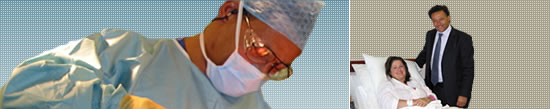
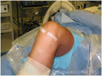
The anaesthetist and Mr. Johnson will see you before surgery. The operation is performed under spinal or general anaesthesia. There will be one or two small incisions in the front of the knee either side of the patellar tendon.
Surgical Technique
Arthroscopic, telescopic, keyhole or minimally invasive surgery is the technique of performing surgery inside a joint through a telescope without disrupting the surrounding structures. Looking inside the joint with the arthroscope allows the surgeon to look directly at the cartilages, ligaments and the articular surfaces without damaging the joint. The arthroscope is a pencil thin tube containing light fibres and is a means of transmitting a picture of the inside of the joint to a video camera. The knee joint is filled with fluid which allows the surgeon to look around the brightly lit inside of the joint. The arthroscope is inserted through a small incision, about 5mm long either side of; and just below the knee cap (patella). As well as the arthroscope a small metal probe isinserted into the knee to help show if there are any tears in the menisci.
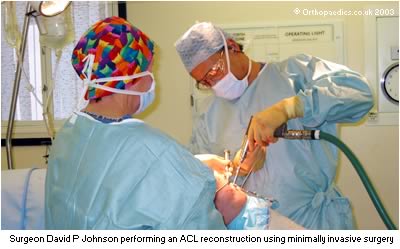
Once extent of the damage to the menisci or other structures in the knee has been determined, small cutting tools are inserted into the joint through the same holes that were used for the arthroscope. With the arthroscope in the joint giving the surgeon a clear view, these small cutting instruments are then used to trim away the damaged part of the meniscus. This is done so that there is once again a smooth surface allowing the joint to roll back and forth without catching or locking. The pieces of meniscus are then removed through the same small holes as the joint is washed out. The fluid is drained out at the end of the procedure, however the knee may feel as if there is a little fluid within it for a few days.
 The arthroscopic technique involves making drill holes through the tibia into the knee joint, and from inside the joint
into the femur. The graft is then passed through these holes so that it lies in the correct position through the joint.
The graft is then secured at each end with a screw.It should also be emphasized that at the time of arthroscopy additional
damage may be found to the cartilages or joint surface. An additional procedure may be undertaken to correct this and this
can usually be done simultaneously without the need for a second operation.
The arthroscopic technique involves making drill holes through the tibia into the knee joint, and from inside the joint
into the femur. The graft is then passed through these holes so that it lies in the correct position through the joint.
The graft is then secured at each end with a screw.It should also be emphasized that at the time of arthroscopy additional
damage may be found to the cartilages or joint surface. An additional procedure may be undertaken to correct this and this
can usually be done simultaneously without the need for a second operation.
Arthroscopy is performed whilst causing as little disruption to the knee as possible, with a minimal of post-operative pain and with a rapid return of function. Patients can generally return to work in a day or two, to sports in a week or two and to normal thereafter. The results and speed of recovery is very much dependant on the nature of the damage to the knee and the extent of surgery undertaken and may as a consequence vary from this norm.
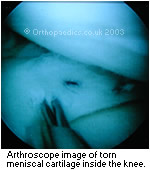
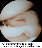 In the case of a tear of the menisci, or "cartilages" of the knee the smooth rolling of the joint surfaces over
the meniscus is prevented as the torn meniscal flap gets caught between the bones of the tibia and the femur. The knee is
usually painful, particularly on twisting, swollen and cannot be fully straightened. Arthroscopic partial meniscectomy is
undertaken to remove the torn piece of meniscus to relieve the pain and swelling, to allow full movement and to prevent
further damage to the knee. The amount of surgery required to remove this torn portion of meniscus will not be known for
certain until the surgeon can see the damage through the arthroscope. However the aim is always to remove as little as the
tear allows.
In the case of a tear of the menisci, or "cartilages" of the knee the smooth rolling of the joint surfaces over
the meniscus is prevented as the torn meniscal flap gets caught between the bones of the tibia and the femur. The knee is
usually painful, particularly on twisting, swollen and cannot be fully straightened. Arthroscopic partial meniscectomy is
undertaken to remove the torn piece of meniscus to relieve the pain and swelling, to allow full movement and to prevent
further damage to the knee. The amount of surgery required to remove this torn portion of meniscus will not be known for
certain until the surgeon can see the damage through the arthroscope. However the aim is always to remove as little as the
tear allows.
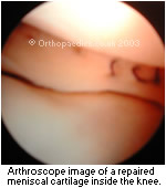
In some cases the meniscus may be able to be repaired. This is where the tear is a "peripheral bucket handle" type, is fairly recent in a patient who usually is under 40 years of age. In this situation Mr. Johnson may be able to suture or stitch the tear edges together through the arthroscope so that it may heal after a period of six weeks. Where possible this may be advantageous in preserving the meniscus and avoiding it's removal. This minimises any potential for excessive knee wear in the future.
 Other arthroscopic procedures commonly undertaken by Mr. Johnson include reconstruction of the knee ligaments by
"arthroscopic anterior cruciate reconstruction", or excision of a fold in the front of the knee by
"arthroscopic excision of a synovial plica", "patellar stabilisation", articular cartilage surgery or
"chondroplasty" or even "osteochondral transplantation" to reconstruct damage to the articular surfaces.
Other arthroscopic procedures commonly undertaken by Mr. Johnson include reconstruction of the knee ligaments by
"arthroscopic anterior cruciate reconstruction", or excision of a fold in the front of the knee by
"arthroscopic excision of a synovial plica", "patellar stabilisation", articular cartilage surgery or
"chondroplasty" or even "osteochondral transplantation" to reconstruct damage to the articular surfaces.
The anaesthetist will see you before surgery. The operation is performed under spinal or general anaesthesia. There will be one or two small incisions in the front of the knee either side of the patellar tendon. A steristrip or a single suture is used to close these wounds.
Following the operation you usually wake up without or with only modest pain because local anaesthetic is inserted into the joint at the end of the procedure. This may wear off after 4 - 6 hours and the tablets to relieve any paid and anti- inflammatory medication is usually prescribed. By the following morning the leg is usually comfortable whilst taking only regular anti-inflammatory medication. Generally only a few hours after your operation you will be able to get around walking without too much difficulty and without crutches. Most arthroscopic surgery patients can be discharged form hospital later the same day as a "Day Case". Sometimes when extensive surgery is performed or operations preformed simultaneously on both legs patients are kept in hospital overnight. After patients go home, they should keep bending and lifting the knee in order to strengthen the muscles and regain movement.
If the knee becomes more swollen over the first week or more painful than during the first day, please return to your general practitioner for early review. Normally you will be reviewed at the hospital by Mr. Johnson after 2 or 3 weeks.
Wound Dressing and Sutures
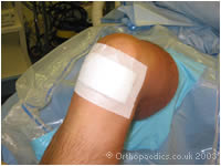
The dressings should be removed after 5 days and the wound inspected. If there is any excessive redness or infection patients should return to the GP or the clinic. If the wounds have been closed with steristrips these may be removed at that time. If sutures were used then the dressings should be maintained for 10 days, following which you should return to the GP's clinic to have the stitches removed. A tubigrip elasticated support should be worn for 7 - 10 days. The wound if dry and the sutures have been removed or dissolved then the wound can be washed, and pool exercises can be begun. If the wound becomes red, inflamed or infected then patients should return to see Mr Johnson.
< BACK to Non-operative Treatment | NEXT: Recovery and Rehabilitation >
Related Links..
+ How to make an appointment
+ Arthroscopy Of The knee - see all links
+ Patient Information Home
+ See the clinic
+ More about Mr Johnson
+ top
© The Bristol Orthopaedics and Sports Injuries Clinic 2003. The Bristol Knee Clinic is a trading name of the Bristol Orthopaedic Clinic Ltd. privacy / copyright | contact | Powered By Create Medical