


Bristol Knee Clinic

| + home | |
| + news | |
| + research | |
| + patient information | |
| + the clinic | |
| + the surgeon | |
| + sport physiotherapy | |
| + sports advice | |
| + medico legal | |
| + products | |
| + resources | |
| + contact | |
| + maps | |
| + directions | |
| + site map |
The Bristol Knee Clinic |
Total Knee Arthroplasty - Surgery
Step-by-step
Quadriceps Exercises
Prior to surgery and post operatively it is important to strengthen the muscles of the leg and to reduce the stiffness. Regular exercises should be undertaken to do this. Static quadriceps exercises consist of tensing the muscle on the front of the thigh whilst the knee is straight. Hold the contraction for 5 to 10 seconds, rest for 5 or 10 seconds and begin again. This should be repeated 10-50 times. Whilst lying on your back the straight leg should be lifted into the air and held for 5 to 10 seconds, then lowered rested for 5 to 10 seconds and repeated 10 to 50 times. Sitting on a high chair or table and bending the knee over the edge will improve knee bending. The good leg may be crossed over the bad one to assist.
Tests and Scans
 Prior to admission to hospital blood tests, a urine test, a knee X-ray, a chest X-ray and an ECG or heart tracing will be
performed. Blood will be cross-matched so as to be available for transfusion following surgery. If you wish to arrange for
your own blood to be pre-donated and ready for autologous transfusion, this should be done some weeks in advance and will
require a trip to the blood bank on several occasions. It is a routine to use blood salvage techniques during and following
surgery, which minimises the need for blood transfusions. Minimally access techniques also reduce the need for blood
transfusions. Prior to the operation any tablets or medications you take, or allergies you may have to medications, should
be brought to the attention of the surgeon. You should stop anti-inflammatory arthritis tablets for one week prior to surgery.
Take only Paracetamol for pain relief during this period. Please notify your surgeon and anaesthetist in advance if you are
taking any anti-coagulants (blood thinners), hormone replacement tablets, the Pill or suffer from diabetes or any other
significant medical condition.
 Surgical technique
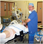
A general anaesthetic is generally used, sometimes a spinal injection is preferred. To be able to completely replace the surface of the knee joint a 15cm incision is made down the front of the knee and the joint is opened.
If a hemi or partial knee replacement is being performed, a smaller incision sometimes of only 5 cm in length may be used.
Minimally Invasive Surgery: MIS
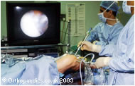 Minimally invasive surgery involves the use of smaller wound incisions and special instrumentation to enable surgery to be
undertaken. These techniques usually result in significant advantages in respect to improve the speed of recovery, speed of
mobilization, shorten hospital stay reduce the period off work and reduce the time until functional and sporting activities
can be resumed. The techniques also usually reduce the amount of post-operative pain experienced and the need for post-
operative pain relief and analgesia. In knee replacement these techniques are applicable and routinely used by Mr. Johnson
in uni-compartmental knee replacement and increasingly in Total Knee Arthroplasty.
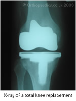 Once the wound has been developed and the knee exposed. The bony overgrowth, which commonly occurs in arthritis of the knee,
is trimmed away and the joint surfaces removed. This involves some shaping of the bone so that the joint replacement components
sit firmly on the bone. The components will either be fixed in situ with acrylic cement or for young patients special components
with a roughened or porous surface will be used. The bone then grows into the roughened surfaces anchoring it.
Once the total knee replacement has been inserted the knee joint is closed over drainage tubes. These tubes take away the bleeding from the knee. They stay in the knee overnight. The knee will have a dressing afterwards and be bandaged. You will have a drip to administer fluids whilst you do not feel like eating or drinking. A blood transfusion may be given if required. Initially the knee may be painful. Powerful pain-killing tablets and injections will be prescribed. It is usual for these to be required regularly for 1 or 2 days and for 1 to 2 weeks intermittently, so do not be afraid to ask if you are in pain. Further blood tests and X-rays will be taken. Injections or tablets to thin the blood and to prevent thrombosis will be given.
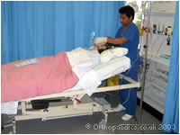 The recovery from the operation requires about 3-7 days in hospital. In this time physiotherapy is commenced. Initially the
leg will be placed on a continuous passive motion machine to gently move the knee through a pre-set range of motion. In
addition, exercises to improve the strength of the quadriceps muscles are performed.
Wound Dressing and Sutures
The crepe bandage, which is applied in theatre, may be removed after 4 days if the wound is satisfactory. The general practitioner or practice nurse should remove the sutures after 14 days. Sometimes arrangements are made for patients to return directly to the hospital for this.
The eight basic steps to undertaking a total knee replacement:
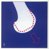
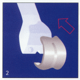
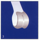
The distal end of the femur is
shaped using a saw to remove
the damaged surfaces and
leave a precice shaped and
sized support for the femoral component which is then fitted.
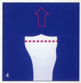
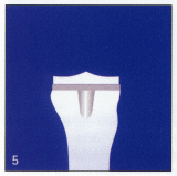
The upper tibia is resected and
the tibial baseplate inserted
into the bone of the upper tibia.
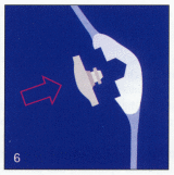
The patellar surface is resected to accept a polyethylene patellar button.
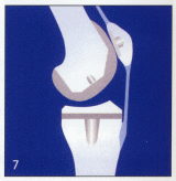
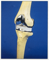
The final knee is assembled
and reduced into position
usually using cement to adhere
the metal
components to the cut
surface of the bones.
< BACK to Non-operative Treatment | NEXT: Recovery and Rehabilitation>
Related Links..
+ How to make an appointment
+ Total Knee Replacement - see all links
+ Patient Information Home
+ See the clinic
+ More about Mr Johnson
+ top
© The Bristol Orthopaedics and Sports Injuries Clinic 2003. The Bristol Knee Clinic is a trading name of the Bristol Orthopaedic Clinic Ltd. privacy / copyright | contact | Powered By Create Medical


