

Bristol Knee Clinic

| + home | |
| + news | |
| + research | |
| + patient information | |
| + the clinic | |
| + the surgeon | |
| + sport physiotherapy | |
| + sports advice | |
| + medico legal | |
| + products | |
| + resources | |
| + contact | |
| + maps | |
| + directions | |
| + site map |
The Bristol Knee Clinic |
Arthroscopy Of The Ankle - Introduction
Ankle Arthroscopy

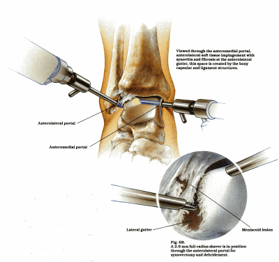
Fig 1. An anterior diagramatic view of an ankle arthroscopy and excision of inflamed synovium from the anterior-lateral
aspect of the ankle joint. This synovitis and impingement is a common problem after ankle sprains.
Our understanding and the development of new surgical techniques have resulted in very significant advances in surgery of the knee and shoulder. In a similar way small joint arthroscopy has resulted in significant advances for elbow, wrist and ankle surgery. Smaller arthroscopes are generally used. The small joints tend to restrict access and free movement within the joint. In the ankle the visualization is improved with distraction of the joint either by a stirrup and weights or by application of a distraction frame. Arthroscopy of these joints are like the knee generally undertaken as a day case procedure with an associated rapid recovery and return to work and sports.
Introduction
Whilst many ankle injuries can be easily diagnosed but some remain difficult to assess with examination and X-ray alone. In less than ten years from the time of the publicaČtion of the first reports on the experience with ankle arthroscopy, the technique has gained increased acceptance in both diagnostic and therapeutic applications. This trend is not surprising. Ankle injuries are frequently seen in orthopaedic practice and, while many are easily diagnosed by clinical and radiographic examination, a number of ankle disorders are either difficult to diagnose or their clinical significance may be difficult to evaluate by traditional methods.
Ankle injuries can result from excessive loading, either as an isolated event (most often a soft tissue injury as the consequence of stress to the inverted foot while in plantar flexion), or as a series of events that produce overuse or fatigue failure. Cartilage and soft tissue injuries associated with recurrent effusion, nonspecific tenderness, restricted motion, or a feeling of instability can present a diagnostic challenge. On the other hand, chondral fractures, osteochondral lesions of the talus, and loose bodies are often more easily identified radiographically, but the extent of articular surface damage may not be readily ascertained. Recent studies and clinical experience have shown that these types of ankle pathologies may be diagnosed and, in many cases, effectively treated, arthroscopically.
< BACK to Arthroscopy Of The Ankle Index | NEXT: Indications / Contra - indications >
Related Links..
+ How to make an appointment
+ Arthroscopy of the Ankle - see all links
+ Patient Information Home
+ See the clinic
+ More about Mr Johnson
+ top
© The Bristol Orthopaedics and Sports Injuries Clinic 2003. The Bristol Knee Clinic is a trading name of the Bristol Orthopaedic Clinic Ltd. privacy / copyright | contact | Powered By Create Medical


