

Bristol Knee Clinic

| + home | |
| + news | |
| + research | |
| + patient information | |
| + the clinic | |
| + the surgeon | |
| + sport physiotherapy | |
| + sports advice | |
| + medico legal | |
| + products | |
| + resources | |
| + contact | |
| + maps | |
| + directions | |
| + site map |
The Bristol Knee Clinic |
Arthroscopy Of The Ankle - Indications / Contra-indications
Anatomy and Portals

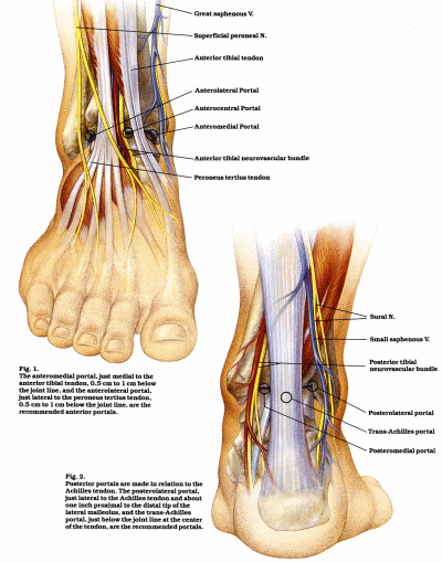
Fig 2: A diagram of the anatomical structures at the anterior and posterior aspect of the ankle joint with the entry sites or
portals marked.

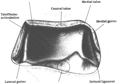
Fig 3: The arthroscopic view of the anterior aspect of the ankle joint.

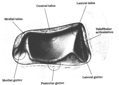
Fig 4: The arthroscopic appearance of the posterior aspect of the ankle joint.

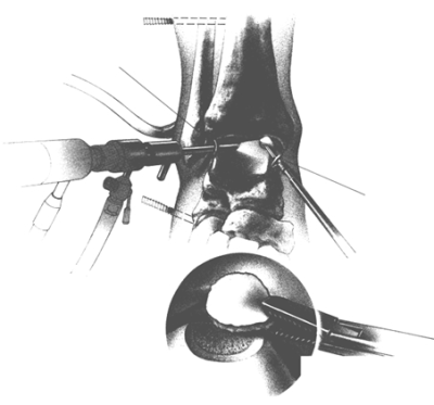
Fig 5: Extraction of a loose fragment of articular cartilage from an osteochondral lesion of the talus.
Who Needs it / Who Doesn't
Patient Selection
Ankle arthroscopy is not presently indicated in as many cases as knee arthroscopy but its value as a relatively non-invasive method of diagnosis and treatment is recognized in a growing number of situations. Diagnostic arthroscopy is indicated in patients whose ankle problems include unexplained pain, swelling, stiffness, instability, hemarthrosis, or locking. Therapeutic ankle arthroscopy is indicated for articular injury, soft tissue and bony impingement, arthrofibrosis, some types of fractures, synovitis, loose bodies and osteophytes, chondromalacia, and osteochondral lesions of the talus. The arthroscopic approach may also be used on occasion for ankle stabilization and arthrodesis.
The only absolute contraindications to ankle arthroscopy are localized soft tissue infection and severe degenerative joint disease. Intra-articular joint infection is not a contraindication and, in fact, can be treated by arthroscopic debridement and drainage. Moderate degenerative joint disease with restricted range of motion is often associated with a reduced joint space, which can prove difficult for access. Severe ankle edema, or a tenuous blood supply may be contraindications to performing arthroscopy in the ankle.
Patient Preparation and Positioning
General, spinal, epidural, or, in some cases, local anesthesia maybe used. Surgeon preference governs patient positioning. Our patients are placed in the supine position with a sandbag supporting the buttock on the operative side. An ankle and a thigh holder, provides several advantages: they facilitate hip, knee, and ankle positioning, permit the surgeon to sit or stand during the procedure, and provide ready access to anterior and posterior portals. A tourniquet is generally used on the thigh.
Key anterior landmarks are used to identify important structures around the ankle. These include the dorsalis pedis artery, saphenous vein, anterior tibial, peroneus tertius and extensor digitorum communis tendons¬. These are marked and avoided. The anterior joint line is located by palpation during dorsiflexion and plantar flexion of the foot. Distraction to increase the space between tibia and talus may then be applied as an optional step, to allow a better visualization. In order to provide complete access to the joint, as well as flexibility of approach during examination and surgery, three portals are routinely established, the anterolateral, the anteromedial, and the posterolateral.
< BACK to Introduction | NEXT: Surgery >
How to arrange an appointment with Mr. Johnson
Your first appointment is usually arranged with Mr Johnson at the Bristol Nuffield Hospital at St Mary's. It is a modern well-equipped hospital with 36 private bedrooms and two operating theatres, and offers a full range of services.
+ How to arrange your first appointment
Related Links..
+ Arthroscopy Of The Ankle - see all links
+ Patient Information Home
+ See the clinic
+ More about Mr Johnson
+ top
© The Bristol Orthopaedics and Sports Injuries Clinic 2003. The Bristol Knee Clinic is a trading name of the Bristol Orthopaedic Clinic Ltd. privacy / copyright | contact | Powered By Create Medical


