

Bristol Knee Clinic

| + home | |
| + news | |
| + research | |
| + patient information | |
| + the clinic | |
| + the surgeon | |
| + sport physiotherapy | |
| + sports advice | |
| + medico legal | |
| + products | |
| + resources | |
| + contact | |
| + maps | |
| + directions | |
| + site map |
The Bristol Knee Clinic |
Arthroscopy Of The Ankle - Surgery
Arthroscopic Surgery of the Ankle
Examination of the Joint
Successful arthroscopic examination of the ankle, like that of the knee or shoulder, requires a methodical approach. With such an approach, the surgeon can be confident that all pathology is visualized, that the method is accurate and reproducible from one patient to another.

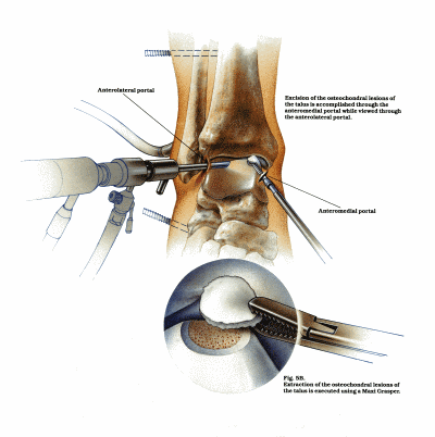
Fig 6:Anterior diagrammatic view of an ankle arthroscopy and removal of a loose fragment of articular cartilage: One of the
most common causes of post traumatic ankle pain.
Ankle Pathology
Osteochondral, arthritic, and soft tissue pathologies in the ankle can be visualized during the arthroscopic examination and many can be treated at once, often without the need for additional open exposure or dissection. Biopsy, debridement, synovectomy and loose body removal procedures can also be performed . Defects of the articular surfaces can be identified. In particular defects of the talus occur commonly. These defects can be identified and often treated.
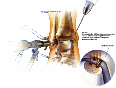
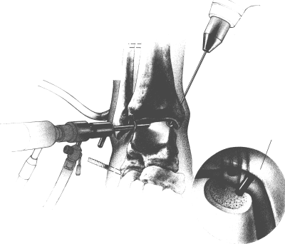
In the past, treatment of such lesions were often associated with delayed diagnosis while significant morbidity and prolonged rehabilitation could be anticipated when arthrotomy was undertaken. Debridement, curettage, and transmalleolar drilling under direct and or fluoroscopic control, to stimulate a new blood supply and healing, can be performed through the arthroscope. The ability to diagnose osteochondral lesions of the talar dome promptly, treat the condition immediately with a relatively non- invasive procedure, and permit early joint motion and patient rehabilitation, are good examples of the advantages offered by ankle arthroscopy.
Arthritic conditions, including loose bodies and osteophytes, are other ankle disorders that can be visualized and treated arthroscopically. Soft tissue pathologies that can also be treated. This included a wide range of synovial disorders, inflammatory conditions, infections, impingement and meniscoid lesions.
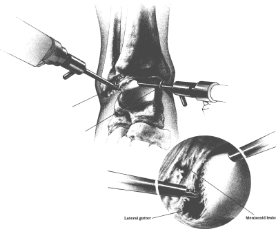
Inversion injuries to the ankle can lead to soft tissue impingement that can cause chronic ankle pain. This soft tissue impingement can be present antero-laterally, postero-laterally or can occur simultaneously in both the antero-lateral and postero-lateral gutters. Soft tissue impingement is most commonly seen in the antero-lateral gutter following torn ankle ligaments.

Fig 7: An anterior diagramatic view of an ankle arthroscopy drilling or fenestration of the subchondral bone in an area
of articular cartilage loss. This promotes the healing of the defect with fibrous tissue.

Fig. 8: Transmalleolar drilling of an osteochondral lesions of the talus through the medial malleolus while viewing through
the anterolateral portal.

Fig. 9: Viewed through the anteromedial portal, anterolateral soft tissue impingement with synovitis and fibrosis at the
anterolateral gutter, this space is created by the bony capsular and ligament structures.
< BACK to Indications / Contra - indications | NEXT: Recovery and Rehabilitation >
Related Links..
+ How to make an appointment
+ Arthroscopy Of The Ankle - see all links
+ Patient Information Home
+ See the clinic
+ More about Mr Johnson
+ top
© The Bristol Orthopaedics and Sports Injuries Clinic 2003. The Bristol Knee Clinic is a trading name of the Bristol Orthopaedic Clinic Ltd. privacy / copyright | contact | Powered By Create Medical


